45 label foot bones
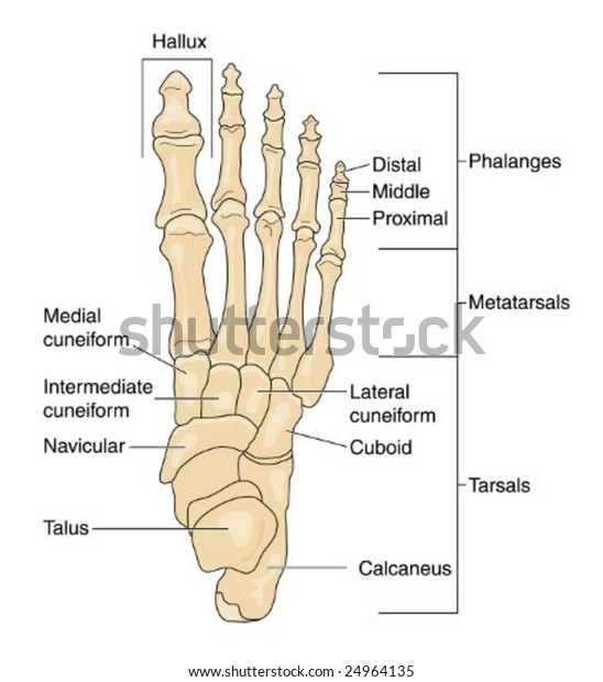
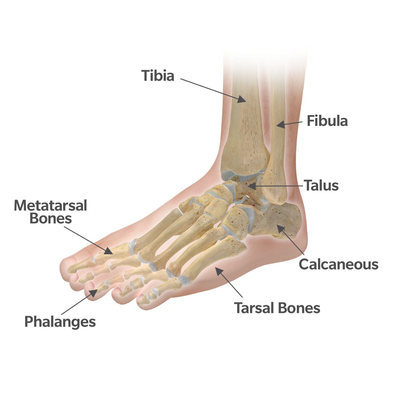
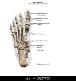
Bones Of The Foot: Feet Anatomy - UMZU There are 26 bones in the human foot. There are three main areas of the foot: the tarsals, metatarsals, and phalanges. These areas are broken down into the bones that they contain: Tarsal - This part of the foot contains 7 bones known as: The talus - attached to the tibia and fibia to help bear the weight of the body. Free Anatomy Quiz - Anatomy of the Foot Bones - Quiz 1 5 - the hand : can you name the bones of the hand? 6 - the appendicular skeleton : learn the bones the arms and legs. 7 - the heart : name the parts of the bones. 8 - the foot : Can you identify the bones of the foot? 9 - the skeleton, posterior : identify the skeleton from behind.
Bones of the Leg and Foot | Interactive Anatomy Guide - Innerbody The bones of the leg and foot form part of the appendicular skeleton that supports the many muscles of the lower limbs. These muscles work together to produce movements such as standing, walking, running, and jumping. At the same time, the bones and joints of the leg and foot must be strong enough to support the body's weight while remaining ...

Label foot bones
Bones of the Foot Diagram - Bodytomy The bones of the foot are divided into anterior region, posterior region, dorsal region, plantar region, distal region, proximal region, medial region, and lateral region. It is important to note, when we say a joint, it is not only about two bones coming together, the muscles, ligaments, and tendons are also a part of it. Foot Bones Labeling Diagram | Quizlet Foot Bones Labeling Diagram | Quizlet Foot Bones Labeling STUDY Learn Write Test PLAY Match Created by maddie_sayatovic Terms in this set (4) Talus ... Calcaneus ... Tarsals ... Metatarsals ... Anatomy of the Foot: Muscles, Tendons, Nerves, and Bones The muscles of the foot are located mainly in the sole of the foot and divided into a central (medial) group and a group on either side (lateral). The muscles at the top of the foot fan out to supply the individual toes. The tendons in the foot are thick bands that connect muscles to bones. When the muscles tighten (contract) they pull on the ...
Label foot bones. Foot (Anatomy): Bones, Ligaments, Muscles, Tendons, Arches and Skin The foot is a part of vertebrate anatomy which serves the purpose of supporting the animal's weight and allowing for locomotion on land. In humans, the foot is one of the most complex structures in the body. It is made up of over 100 moving parts - bones, muscles, tendons, and ligaments designed to allow the foot to balance the body's ... Label the Foot Bones - Printable - PurposeGames.com This is a printable worksheet made from a PurposeGames Quiz. To play the game online, visit Label the Foot Bones Download Printable Worksheet Please note! You can modify the printable worksheet to your liking before downloading. Download Worksheet Include correct answers on separate page About this Worksheet Skeletal System - Labeled Diagrams of the Human Skeleton - Innerbody Capitate Bone Distal Phalanges of the Hand Hamate Bone Lunate Bone Middle Phalanges of the Hand Palmar Carpometacarpal Ligaments Pisometacarpal Ligament Proximal Phalanges of the Hand Scaphoid Bone Trapezium Bone Trapezoid Bone Triquetral Bone (Triquetrum) LEG AND FOOT Femur Hip Joint Replacement Oblique Popliteal Ligament Patella Patellar Ligament Bones Of Foot Anatomy, Function & Diagram | Body Maps - Healthline The 26 bones of the foot consist of eight distinct types, including the tarsals, metatarsals, phalanges, cuneiforms, talus, navicular, and cuboid bones. The skeletal structure of the foot is...
Bones of the Foot - Tarsals - Metatarsals - TeachMeAnatomy The talus is the most superior of the tarsal bones. It transmits the weight of the entire body to the foot. It has three articulations: Superiorly - ankle joint - between the talus and the bones of the leg (the tibia and fibula). Inferiorly - subtalar joint - between the talus and calcaneus. Foot Labeling Quiz - Sheridan College Lateral Right ... Return to Menu Foot Diagram: Labeled Anatomy | Science Trends There are 5 metatarsal bones in the human foot: First metatarsal bone Second metatarsal bone Third metatarsal bone Fourth metatarsal bone Fifth metatarsal bone The first metatarsal bone is the thickest and shortest of all the metatarsals, while the second metatarsal bone is the longest of them all. Foot bones: Anatomy, conditions, and more - Medical News Today The talus, or ankle bone: The talus is the bone at the top of the foot. It connects with the tibia and fibula bones of the lower leg. The calcaneus, or heel bone: The calcaneus is largest of the ...
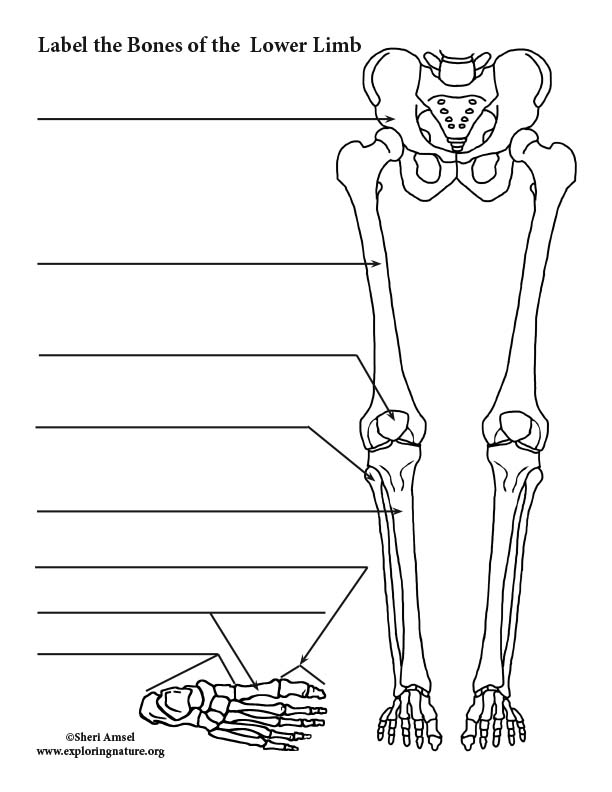
Leg Bones Anatomy, Names & Diagram | Leg & Foot Bones - Video & Lesson ... Femur: the upper leg in both legs. Patella: the kneecap in both legs. Tibia: the larger of the two bones in the lower leg, which supports the body. Fibula: the smaller bone in the lower leg, which ... Foot x-ray - labeling questions | Radiology Case - Radiopaedia Lesser metatarsal sesamoid bone (of fifth metatarsal) 28. Second metatarsophalangeal joint. 29. Proximal phalanx third toe. 30. Proximal interphalangeal joint third toe. 31. Middle phalanx second toe ... Foot oblique labeled. 1. Lateral malleolus. 2. Medial malleolus. 3. Tibiotalar joint (ankle joint) 4. Superior facet of trochlea of talus. 5 ... GetBodySmart | Interactive Anatomy and Physiology GetBodySmart | Interactive Anatomy and Physiology foot bone labeled foot bone labeled Pin by jeremy enfinger on nursing. Print hesi flashcards. Talus anatomy bone left body surface inferior articular talar below bones calcaneal tarsal facet posterior ankle foot human articulates tarsus foot bone labeled
PDF Bone Labeling Exercise - Mesa Community College LABELING EXERCISE: BONES OF THE AXIAL AND APPENDICULAR SKELETON . Most, but not all, features you are required to know are shown on the following pages. Study from the bone list or your textbook after you marked the drawings as instructed on page 6-2. After you have studied the bones in lab, label the drawings as a self-test. Do not spend your
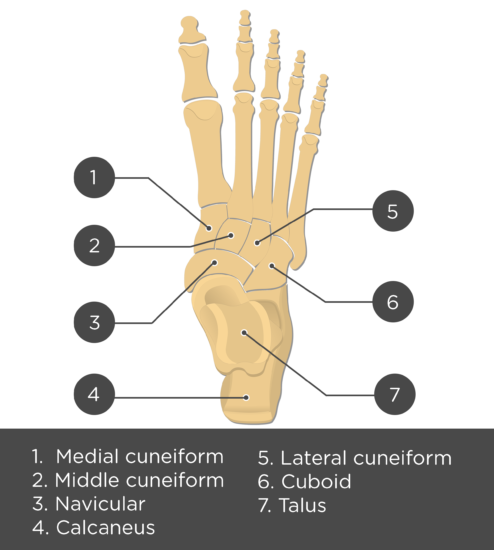
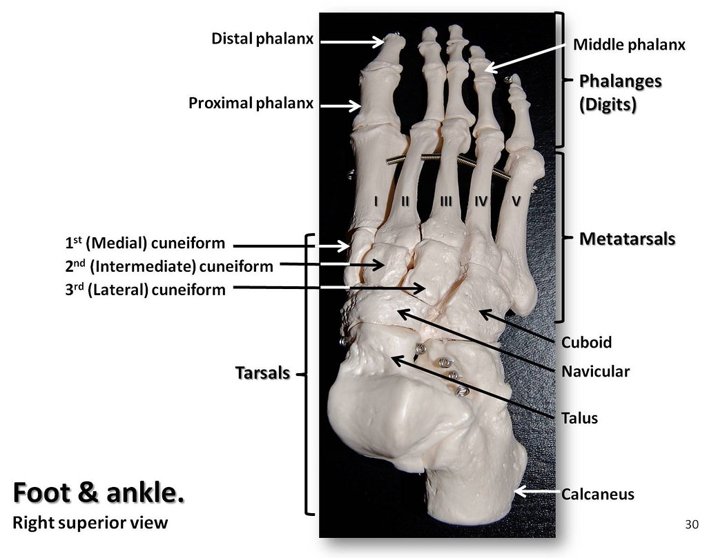
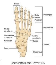
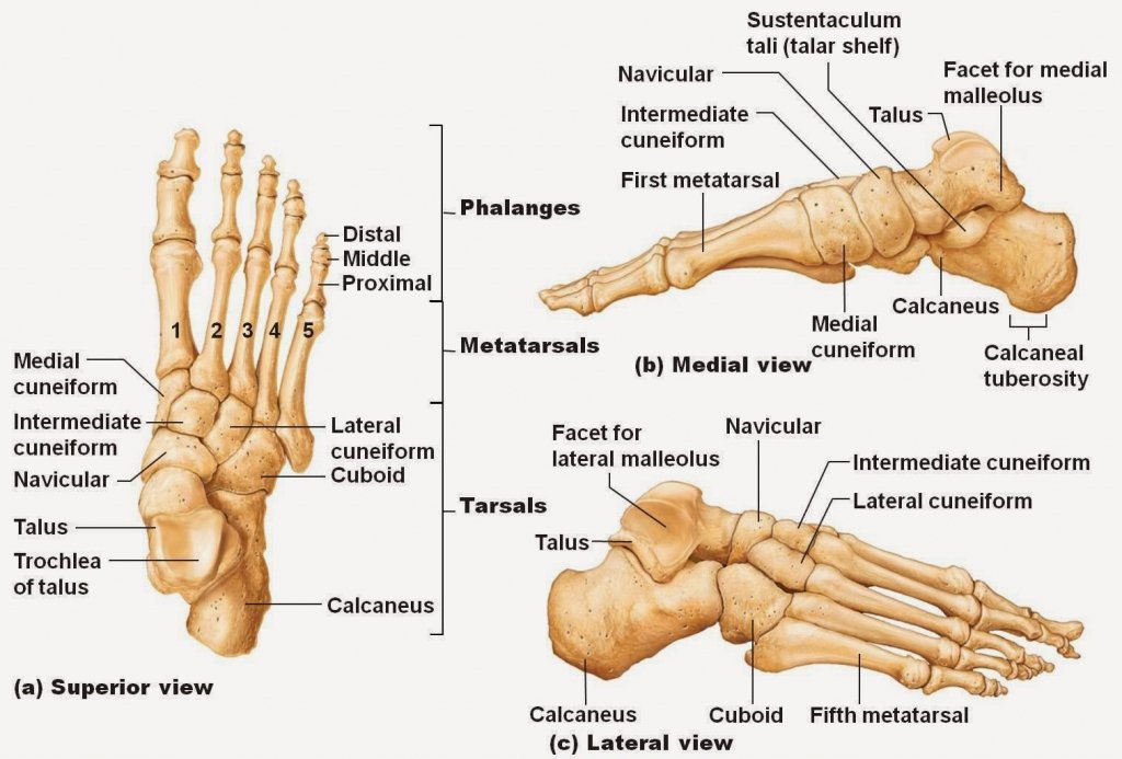
Foot and Ankle Anatomy: Tarsal Bone Mnemonic - EZmed Foot and Ankle Anatomy There are 7 tarsal bones in the foot: calcaneus, talus, navicular, cuboid, medial cuneiform, intermediate cuneiform, and lateral cuneiform. By definition, the tarsal bones function to articulate with the tibia and fibula proximally and the metatarsals distally to form the ankle joint, hindfoot, and midfoot.
Label the Foot Bones Quiz - PurposeGames.com This is an online quiz called Label the Foot Bones There is a printable worksheet available for download here so you can take the quiz with pen and paper. Your Skills & Rank Total Points 0 Get started! Today's Rank -- 0 Today 's Points One of us! Game Points 11 You need to get 100% to score the 11 points available Actions Image Quiz Image Quiz
bones of foot labeled - Microsoft bones foot lower limb anatomy left lateral medial right bottom superior posterior tarsal figure metatarsal toes mid phalanges physiology divided Diagram Showing Parts Of The Foot underside nerves ggxx5 Bones Of Foot foot bones anatomy netter pricing labeled Foot Bones: Anatomy & Injuries - Foot Pain Explored
Foot Bones: Anatomy & Injuries - Foot Pain Explored The talus is the highest foot bone. It forms the bottom of the ankle joint, articulating with the tibia and fibula (shin bones) and the top of the subtalar joint, articulating with the calcaneus (heel bone). Interestingly, no muscles attach to the talus. The talus is held in place by the foot bones surrounding it and various ligaments. Calcaneus
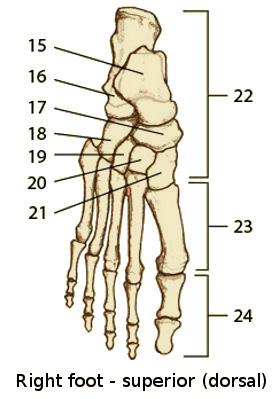
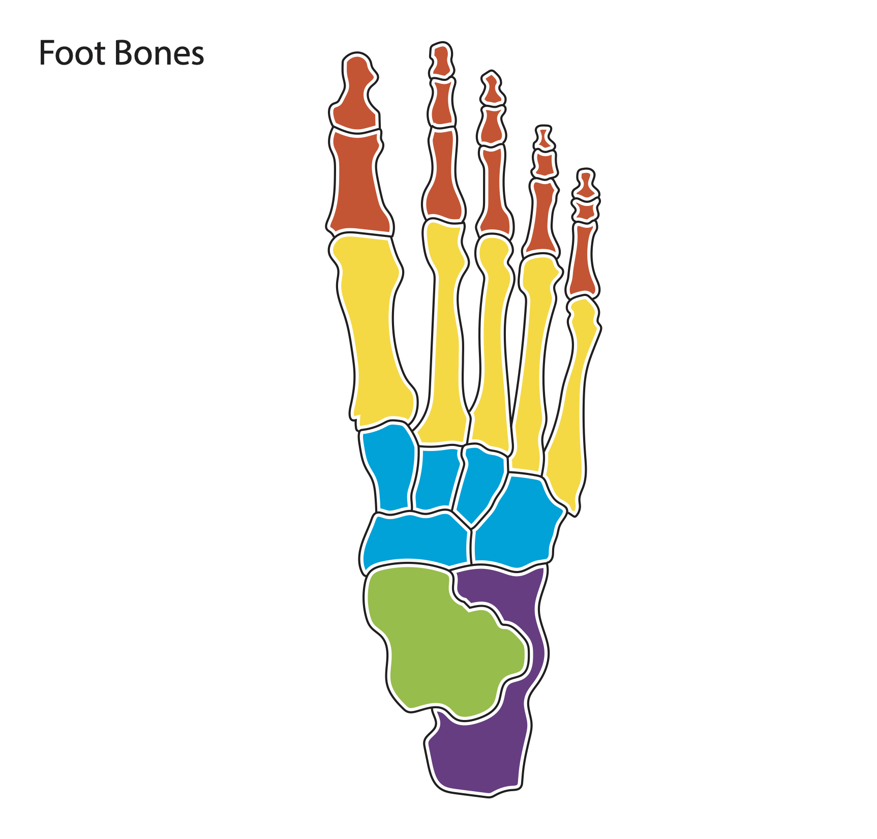
Foot Bones, Parts, Skeleton, Pictures (All You Need To Know) - MEDPLUX The foot bones are generally grouped into tarsal, metatarsal and phalanges. So, to simplify, the hindfoot and midfoot consist of 7 tarsal bones (calcaneus, talus, navicular, cuboid, and 3 cuneiforms) while the forefoot consist of 5 metatarsal bones and 14 phalanges. Hindfoot Bones Anatomy foot skeleton picture
Label The Structures Of The Ankle And Foot - Royal Pitch The structure of the foot and ankle is composed of bones, joints, tendons, and muscles. The tibia and fibula form the midfoot and forefoot, respectively. The talus is the biggest bone in the foot and forms the hindfoot. Its three protrusions are called the tibia and the talus. The talus is connected to the calcaneus at the subtalar joint.
Foot bones: label Diagram | Quizlet Start studying Foot bones: label. Learn vocabulary, terms, and more with flashcards, games, and other study tools.
Foot Bones Anatomy and Mnemonic - Registered Nurse RN Metatarsal Bones (Metatarsus) of the Foot. Proximal to the phalanges are the five metatarsal bones, which together make up the metatarsus of the foot. The prefix "meta" means "beyond or after.". They are beyond the tarsals, which I'll cover in a moment. Again, these bones are numbered 1-5, with one being on the side of your big toe ...
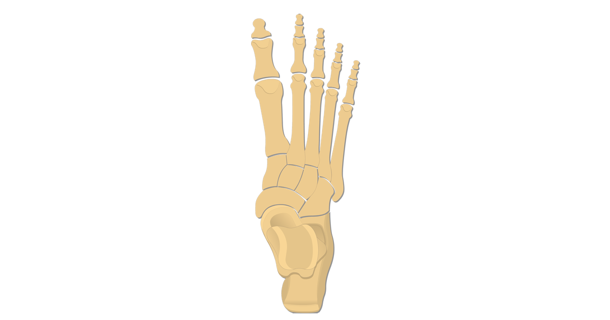
Bones of the Foot Quiz Anatomy - Registered Nurse RN You will be required to label the cuboid, navicular, calcaneus, lateral cuneiform, medial cuneiform, medial cuneiform, talus, metatarsals, and distal/middle/proximal phalanges.j If you have not yet reviewed this material, you can review our foot bones anatomy notes. In addition, you can view our foot bones anatomy video on YouTube.
The Human Skeleton: All You Need to Know - Bodytomy The calf bone or fibula is the smaller of the two bones that form the lower leg. It is placed laterally to tibia and is the most slender of all the long bones. Tarsus. The tarsus or heel bone consist of 7 bones that make up the posterior part of the foot, that is present between the tibia, fibula and metatarsals.
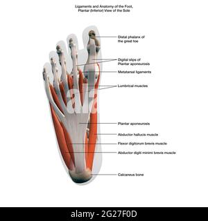
Anatomy of the Foot: Muscles, Tendons, Nerves, and Bones The muscles of the foot are located mainly in the sole of the foot and divided into a central (medial) group and a group on either side (lateral). The muscles at the top of the foot fan out to supply the individual toes. The tendons in the foot are thick bands that connect muscles to bones. When the muscles tighten (contract) they pull on the ...
Foot Bones Labeling Diagram | Quizlet Foot Bones Labeling Diagram | Quizlet Foot Bones Labeling STUDY Learn Write Test PLAY Match Created by maddie_sayatovic Terms in this set (4) Talus ... Calcaneus ... Tarsals ... Metatarsals ...
Bones of the Foot Diagram - Bodytomy The bones of the foot are divided into anterior region, posterior region, dorsal region, plantar region, distal region, proximal region, medial region, and lateral region. It is important to note, when we say a joint, it is not only about two bones coming together, the muscles, ligaments, and tendons are also a part of it.














Post a Comment for "45 label foot bones"