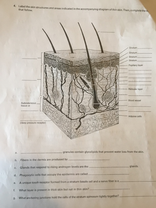
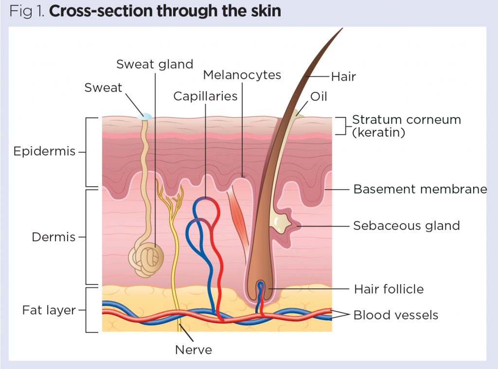
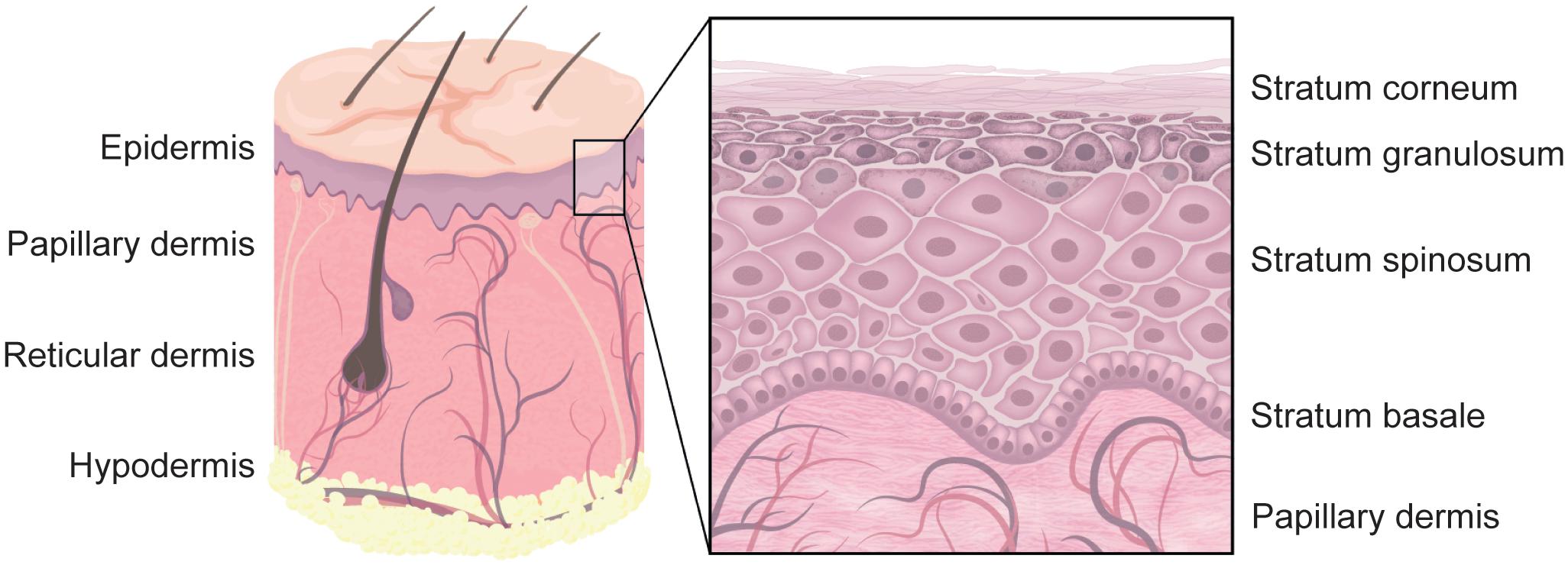
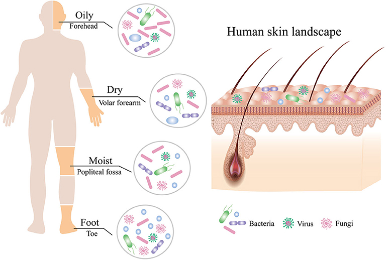
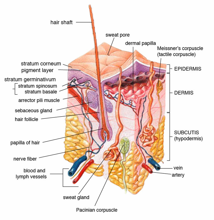
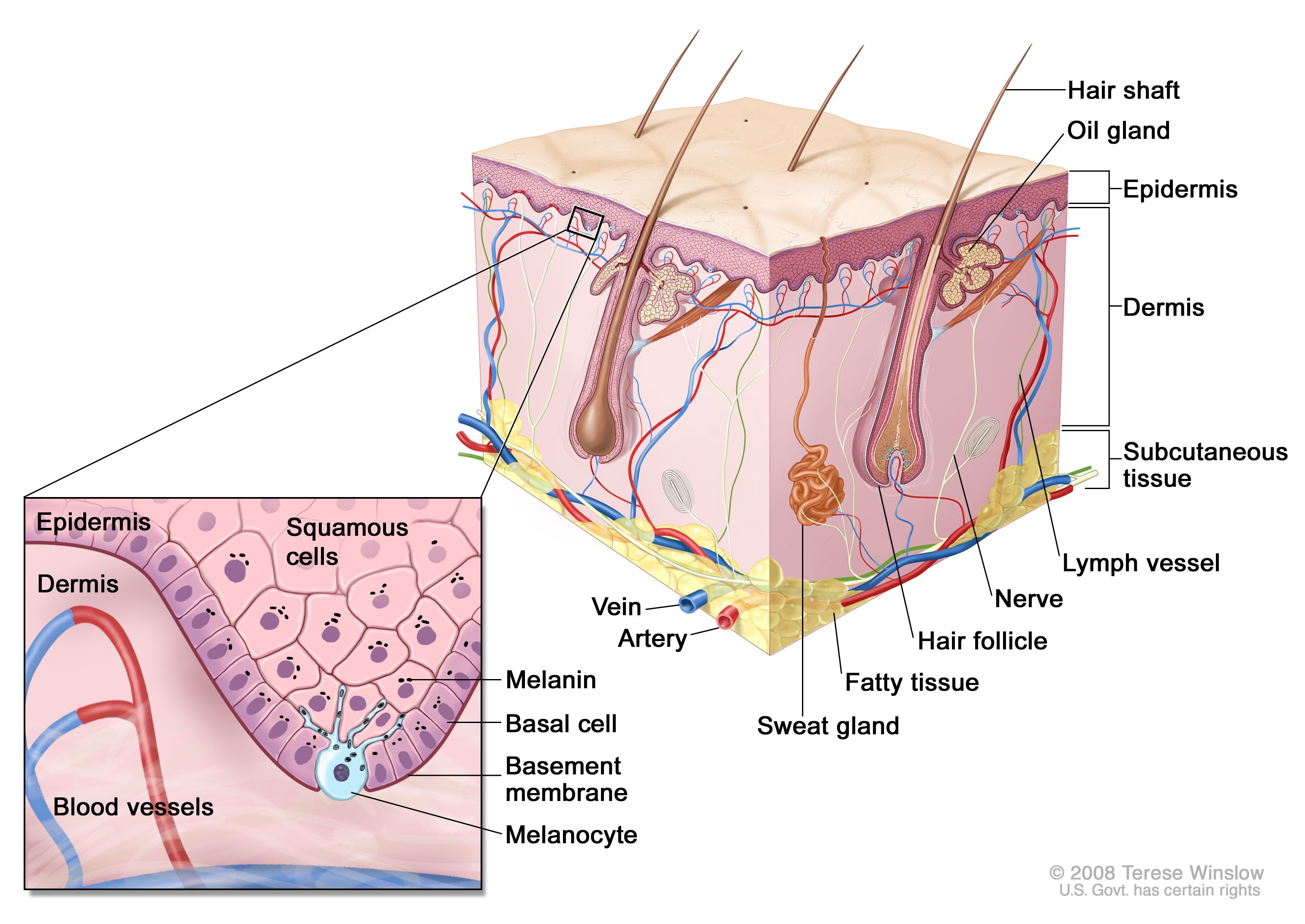
40 label the skin structure and areas indicated in the accompanying diagram of skin
Chernobyl disaster - Wikipedia The Chernobyl disaster (also called the Chornobyl disaster) was a nuclear accident that occurred on 26 April 1986 at the No. 4 reactor in the Chernobyl Nuclear Power Plant, near the city of Pripyat in the north of the Ukrainian SSR in the Soviet Union. It is one of only two nuclear energy accidents rated at seven—the maximum severity—on the International Nuclear Event Scale, … Success Essays - Assisting students with assignments online Get 24⁄7 customer support help when you place a homework help service order with us. We will guide you on how to place your essay help, proofreading and editing your draft – fixing the grammar, spelling, or formatting of your paper easily and cheaply.
PDF Label The Skin Structures And Areas Indicated - Paris Saint-Germain F.C. red blood cells circulating in the dermal capillaries and, skin structure diagram to label is free hd wallpaper this wallpaper was upload at march 10 2017 upload by admin in structure body, label the skin structures and areas indicated in the accompanying diagram of thin skin then complete the
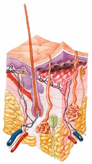
Label the skin structure and areas indicated in the accompanying diagram of skin
Label the Skin | Biology Diagram | Quizlet Epidermis. layer of the skin that is continually being shed to make room for new skin. Dermis. layer of the skin that contains nerves, vessels, hair follicles, glands, and muscles. Hypodermis. layer of the skin that is used for fat storage. Sweat Pore. opening in the skin that sweat comes out of. Hair Follicle. PDF Skin Diagram Labeling - New Providence School District Skin Diagram Labeling . 1. Label the diagram with the . letters. below according to the structure/area they describe. You may label with a line or put the label directly onto the area described. Be as precise as possible. If you are worried about the precision of your label add a word after to explain exactly where your label should be. Layers of the Skin | Anatomy and Physiology I - Lumen Learning The skin is composed of two main layers: the epidermis, made of closely packed epithelial cells, and the dermis, made of dense, irregular connective tissue that houses blood vessels, hair follicles, sweat glands, and other structures. Beneath the dermis lies the hypodermis, which is composed mainly of loose connective and fatty tissues.
Label the skin structure and areas indicated in the accompanying diagram of skin. ACS Applied Materials & Interfaces | Vol 12, No 12 Mar 25, 2020 · A novel double-helical metal–organic framework (dhMOF) was constructed using a butterfly-shaped, electron-rich π-extended tetrathiafulvalene ligand containing four benzoate groups (ExTTFTB). Iodine-mediated oxidation of half of the ExTTFTB ligands, possibly one in each loop, created π donor–acceptor chains along the seams of neighboring double helices, … Label Skin Diagram Printout - EnchantedLearning.com epidermis - the outer layer of the skin. hair follicle - a tube-shaped sheath that surrounds the part of the hair that is under the skin. It is located in the epidermis and the dermis. The hair is nourished by the follicle at its base (this is also where the hair grows). hair shaft - The part of the hair that is above the skin. Solved tive tissue 4. Label the skin structures and areas - Chegg Label the skin structures and areas indicated in the accompanying diagram of thin skin. Then, complete the statements that follow. Weisshaft Stratum opidamist Stratum Stratum Stratum Papilary layer Dermis Reticular layer ascectors allmusde Encrine Sweet blond Dermal Vascular plexus pensery neare fiber Blood vessel Subcutaneous tissue or Assignment 11 pg 104.pdf - 4. Label the skin structures and areas ... Label the skin structures and areas indicated in the accompanying diagram of thin skin. Then, complete the statements that follow. Subcutaneous J tissue or _l T~P-r
The Integumentary System_page2_answers.png - 4. Label the skin ... View The Integumentary System_page2_answers.png from BIO 1012 at South University, Savannah. 4. Label the skin structures and areas indicated in the accompanying diagram of thin skin. Then, complete Essay Fountain - 24/7 Professional Care about Your Writing We offer essay help for more than 80 subject areas. You can get help on any level of study from high school, certificate, diploma, degree, masters, and Ph.D. some of the subject areas we offer assignment help are as follows: Clinical Instructions for Using Silver Diamine Fluoride (SDF) in … Condition the lesion and surrounding areas with 20% polyacrylic acid for 10 seconds (removing the smear layer and activating the surface for ionic exchange). It is important to condition not just the lesion but the surrounding areas as well. 5. Rinse with water for 10 seconds and blot dry (leaving a moist "glossy" surface). 6. (PDF) Nanda NIC NOC | dwi adiyanto - Academia.edu Enter the email address you signed up with and we'll email you a reset link.
Skin Diagram with Detailed Illustrations and Clear Labels - BYJUS Skin Diagram. The largest organ in the human body is the skin, covering a total area of about 1.8 square meters. The skin is tasked with protecting our body from external elements as well as microbes. The skin is also responsible for maintaining our body temperature - this was apparent in victims who were subjected to the medieval torture of ... Solved 4. Label the skin structures and areas indicated in | Chegg.com 4. Label the skin structures and areas indicated in the accompanying diagram of thin skin. Then, complete the that follow Stratum Stratum Stratum. Stratum Popilary layer Reticular layer Blood vessel tissue or Adpose cels (deep pressure receptor granules contain glycolipids that prevent water loss from the skin. b. Fibers in the dermis are ... Skin Structure (Labeling) Flashcards | Quizlet Start studying Skin Structure (Labeling). Learn vocabulary, terms, and more with flashcards, games, and other study tools. Braun Series 9 Model Comparison: What Are The Differences? See the price on Amazon. The updated Series 9 shavers (first introduced in 2016) use the 92xx template for the model names: 9290cc, 9295cc, 9297cc, 9293s, 9260s, etc.. They come with an updated shaving head (cassette) and all the models are suitable for wet & dry use.
Neufert 4th Edition | PDF | Elevator | Roof - Scribd Ernst and Peter Neufert. llliii. I Fourth Edition. Updated by Professor Johannes Kister on behalf of the Neufert Foundation with support from the University of Anhalt Dessau Bauhaus (Dipl. lng. Mathias Brockhaus, Dipl. lng. Matthias Lohmann and Dipl. lng. Patricia Merkel). TRANSLATED BY DAVID STURGE (5BWILEY-BLACKWELL A John Wiley & Sons, Ltd., Publication English …
Anatomy and Physiology 2e - 2e - Open Textbook Library Anatomy and Physiology 2e is developed to meet the scope and sequence for a two-semester human anatomy and physiology course for life science and allied health majors. The book is organized by body systems. The revision focuses on inclusive and equitable instruction and includes new student support. Illustrations have been extensively revised to be clearer and …
Layers of the Skin | Anatomy and Physiology I - Lumen Learning The skin is composed of two main layers: the epidermis, made of closely packed epithelial cells, and the dermis, made of dense, irregular connective tissue that houses blood vessels, hair follicles, sweat glands, and other structures. Beneath the dermis lies the hypodermis, which is composed mainly of loose connective and fatty tissues.
PDF Skin Diagram Labeling - New Providence School District Skin Diagram Labeling . 1. Label the diagram with the . letters. below according to the structure/area they describe. You may label with a line or put the label directly onto the area described. Be as precise as possible. If you are worried about the precision of your label add a word after to explain exactly where your label should be.
Label the Skin | Biology Diagram | Quizlet Epidermis. layer of the skin that is continually being shed to make room for new skin. Dermis. layer of the skin that contains nerves, vessels, hair follicles, glands, and muscles. Hypodermis. layer of the skin that is used for fat storage. Sweat Pore. opening in the skin that sweat comes out of. Hair Follicle.












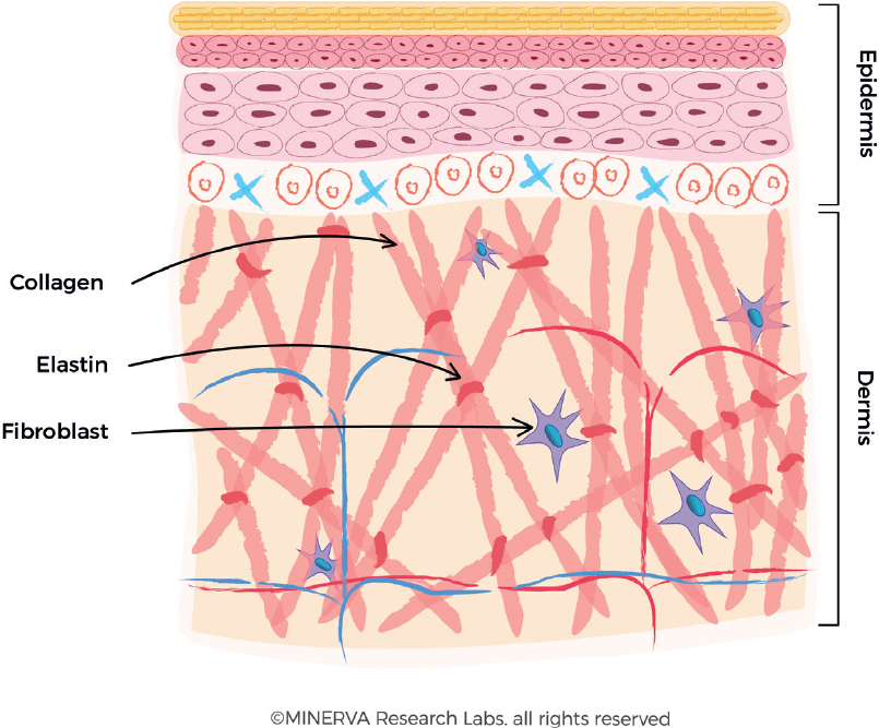

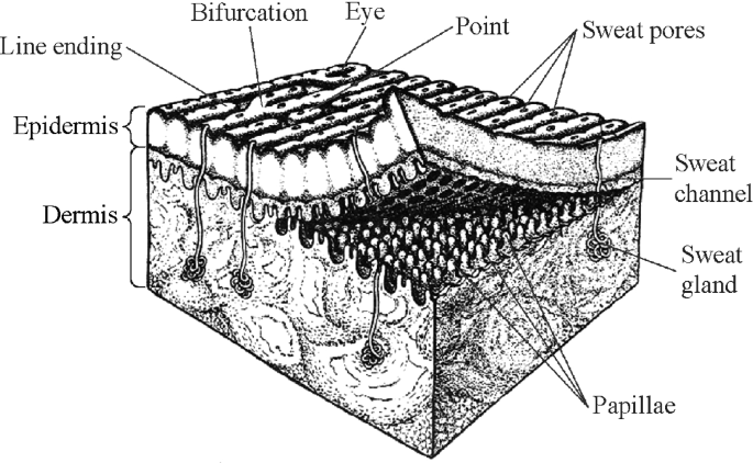


Post a Comment for "40 label the skin structure and areas indicated in the accompanying diagram of skin"