42 art-labeling activity: neuron structure
A Labelled Diagram Of Neuron with Detailed Explanations Diagram Of Neuron. A neuron is a specialized cell, primarily involved in transmitting information through electrical and chemical signals. They are found in the brain, spinal cord and the peripheral nerves. A neuron is also known as the nerve cell. The structure of a neuron varies with their shape and size and it mainly depends upon their ... Answered: Art-labeling Activity: Structure of a… | bartleby Art-labeling Activity: Structure of a lymph node Medulla (B cells Cortex (B cells) and macrophages) Medullary sinus Medullary cord Paracortex (T cells) Efferent vessel Capsule Hilum Subcapsular Afferent vessel space Lymph node artery and vein Trabeculae • Previous Question Drag the labels to the appropriate location in the figure. fullscreen Expand
Answered: Chapter 27 Homework - Altempt… | bartleby Question. Transcribed Image Text:

Art-labeling activity: neuron structure
Best chapter 4 adaptive follow-up Flashcards - Quizlet Art-labeling Activity: Neuron structure PICTURE Reading Quiz - Chapter 4 Question 7 Which connective tissue type is found in the walls of large blood vessels and in ligaments supporting transitional epithelia? a) dense irregular connective tissue proper b) elastic connective tissue proper c) adipose connective tissue proper Neuron Labeling Teaching Resources | Teachers Pay Teachers Digitally interactive drag and drop labeling activity to learn the structures of the brain and neuron. This reviews the structure, function and information processing done by the lobes and parts of the brain along with the parts of a nerve cell. Solved Art-labeling Activity: Neuron Structure 6 of 36 - Chegg Art-labeling Activity: Neuron Structure 6 of 36 Review Part A Drag the labels to the appropriate location in the figure. Axon Nucleus Synaptic terminals Microfibrils and microtubules CO on Coll body II. Mitochondrion Dendrites Nucleolus Submit Request Answer Solv cheo
Art-labeling activity: neuron structure. PDF Nervous System Characteristics - Napa Valley College Figure 13.3 A Review of Neuron Structure Dendrites Cell body Axon Terminal boutons Stimulated by environmental changes or (action potential) toward the activities of other cells Contains the nucleus, mitochondria, ribosomes, and other organelles and inclusions Conducts nerve impulse synaptic terminals Affect another neuron or effector organ Solved Art-Labeling Activity: Structure of nervous tissue - Chegg Expert Answer 100% (12 ratings) 1 - Nucleus 2- Axon 3- Nucleolus 4- Dendrites 5- Neuroglial cells 6 - ECM ( Extracellular matrix) 7- Cell body of neuron Nervous tissue consists of neurons and Neuroglial cells. Neurons are the structural … View the full answer Answered: Art-labeling Activity: Structural… | bartleby match to correct box. Transcribed Image Text: Art-labeling Activity: Structural organization of skeletal muscle Reset Help Epimysium Muscle fascicle Endomysium Perimysium Nerve Muscle fibers Blood vessels Tendon Muscle fiber (cell) Week 4 Chapter 13_.pdf - Week 4 Chapter 13_ Due: 11:59pm ... - Course Hero ANSWER: Correct Art-labeling activity: Histology of neural tissue in the CNS A neuron in the cerebrum of the brain sends an impulse to the cerebellum of the brain. A neuron carries information from the stomach area to the brain. A neuron in the spinal cord sends an impulse to the upper limb, removing the hand from a hot object.
PDF Screen Shot 2019-03-23 at 10.52.26 AM - Los Angeles Mission College Activity 3 Studying the Structure of a Neuromuscular Junction 1. If possible, examine a three-dimensiona model of skeletal ... Label the axon of the motor neuron, its terminal branches, axon termi- na s, and muscle fibers. ron arcolemma ... figure as an Art Labeling Activity Mastering using Motor neuron axon branches Terminal branch of an axon Neural Tissue > Chapter Quizzes > Art-labeling Activities Label the following structural components of a neuron. dendrite - C Mitochondrion - B Nissl bodies (RER and free ribosomes) - A telodendria - E synaptic terminals - F axon - D Correctly label the cells of the central nervous system on the diagram. neuron(s) - A ependymal cell - D oligodendrocyte - B microglial cell - C astrocyte - E Art labeling Activity Figure 4.3.png - | Course Hero Exercise 4 Review Sheet Art labeling Activity 1 2 of 2.png. 1. Exercise 4 Review Sheet Reading Question 3.png. Coastal Carolina Community College. ... If the sulfur dichloride molecule,SCl2, were to form, what would its structure look like? Use a bond (−−−−−−) to represent a bonding pair of electrons and two dots (∙∙ ) to ... Answer correct art based question chapter 4 question - Course Hero ANSWER: Correct Neuroglial cells have many functions, including anchoring neurons and blood vessels in place, monitoring the composition of the extracellular fluid, speeding up the rate of nerve impulse transmission, and circulating the fluid that surrounds the brain and spinal cord.
Chapter 12 - Central Nervous System (CNS) Flashcards - Quizlet The brain and spinal cord compose the __________. central nervous system. The autonomic nervous system does NOT carry signals to: skeletal muscle. Art-Labeling Activity: Neuron Structure. Art-Labeling Activity: The Myelin Sheath in the PNS and CNS. Art-Labeling Activity: Neuroglial Cells of the CNS. Ch11 HW- Introduction to the Nervous System and Nervous ... - Course Hero Dendrites are branched extensions off of a neuron. Neuroglia are the supporting cells of the nervous system. ... ANSWER: somatic motor division central nervous system peripheral nervous system Correct ArtLabeling Activity: Neuron Structure Part A Drag the appropriate labels to their respective targets. Neuron Labelling Teaching Resources | Teachers Pay Teachers You can use this resource to label parts of a neuron as a model, diagram, or notes in your nervous system unit to instruct, explain, and facilitate student learning about neurons' design in the human body. Science from the South Doodle Docs can be used as a part of many different activities for your students, such as notes, part of an interactive n Nervous System Structure and Function Labeling and Coloring Activities ... Nervous System Structure and Function Labeling and Coloring Activities - Downloadable Only. Price: $6.95. Add to Cart. View Cart. This bundle includes 14 Nervous System anatomy assessment activities for high school and college anatomy students, including: diagram labeling and coloring pages. All answer keys included. (28 pages total).

Ch 13_lecture_presentation
Diagram Quiz on Neuron Structure and Function (Labeling Quiz) Diagram Quiz on DNA replication. 1. Identify the cell type in the above figure. 2. In the figure, labeled '1' receives impulses from adjacent neuron. It is called the. 3. In the figure, labeled '2' is the short filaments from the cell body that carries impulses from dendrites to the cell body which is the. 4.

Drag The Labels Onto The Diagram To Identify The Various Synapse ...
art labeling activity figure 14.9 - dlicioustastytoo Art Labeling Activity. Label the nerves on the Lumbar Plexus. Records the start and stop times the activity label around these times may not correspond to the actual activity per-formed. In some they converge to a narrow attachment Figure 10-4B and in some they are oblique and pennate Figure 10-4C like the feathers in an old-fashioned plume.
35 Neuron Labeled Diagram - Wiring Diagram Database
A&P Ch.11-13 Lecture Test Flashcards - Quizlet Art-Labeling Activity: Neuroglial Cells of the CNS The small phagocytic cells that engulf debris and pathogens in the CNS are the __________. microglia (smallest/ least abundant) The neurotransmitter involved in emotion, motivation, and addictive behavior is __________. dopamine

33 Brain Anatomy Quiz Label - Labels Database 2020
Label a Neuron Quiz - PurposeGames.com Latest Activities. An unregistered player played the game 1 hour ago; An unregistered player played the game 1 hour ... About this Quiz. This is an online quiz called Label a Neuron. There is a printable worksheet available for download here so you can take the quiz with pen and paper. From the quiz author. Title says it all. Your Skills & Rank ...

Exam 2, Chapter 12, diagrams labeling Flashcards | Quizlet
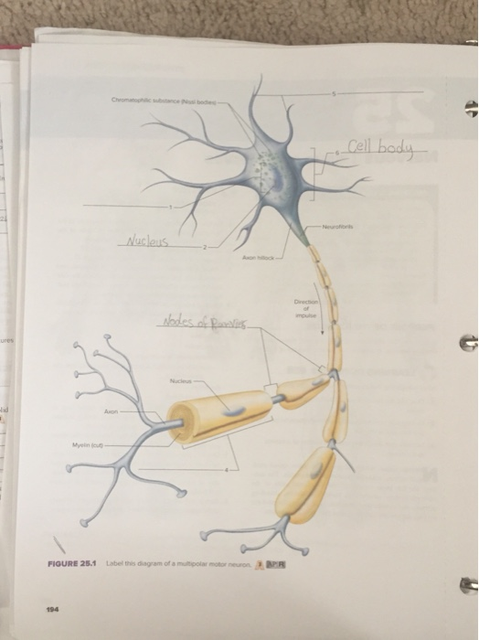
Ch 12 Flashcards | Quizlet Art-labeling Activity: Structure of a typical motor neuron (2 of 2) Which factors contribute to increasing the speed of nerve impulse transmission? larger diameter of axon and the presence of myelin sheath Which of the following statements describes interneurons?
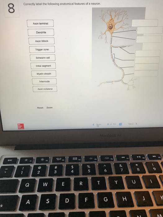
32 Correctly Label The Following Anatomical Features Of A Neuron ...
Kami Export - Esmeralda Castillo Derma - Exercise 15 HIstology of ... Instructors may assign this figure as an Art Labeling Activity using Mastering A&PTM Capillary Neuron Astrocyte (a) Astrocytes are the most abundant CNS neuroglia. (b) Microglial cells are defensive cells in the CNS. Fluid-filled cavity Neuron Microglial cell Cilia Ependymal cells Brain or spinal cord tissue (c) Ependymal cells line ...

Human physiology
Solved Art-labeling Activity: Neuron Structure 6 of 36 - Chegg Art-labeling Activity: Neuron Structure 6 of 36 Review Part A Drag the labels to the appropriate location in the figure. Axon Nucleus Synaptic terminals Microfibrils and microtubules CO on Coll body II. Mitochondrion Dendrites Nucleolus Submit Request Answer Solv cheo
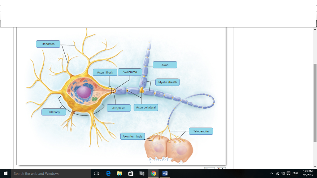
Neuron Diagram Labeled - Human Anatomy
Neuron Labeling Teaching Resources | Teachers Pay Teachers Digitally interactive drag and drop labeling activity to learn the structures of the brain and neuron. This reviews the structure, function and information processing done by the lobes and parts of the brain along with the parts of a nerve cell.
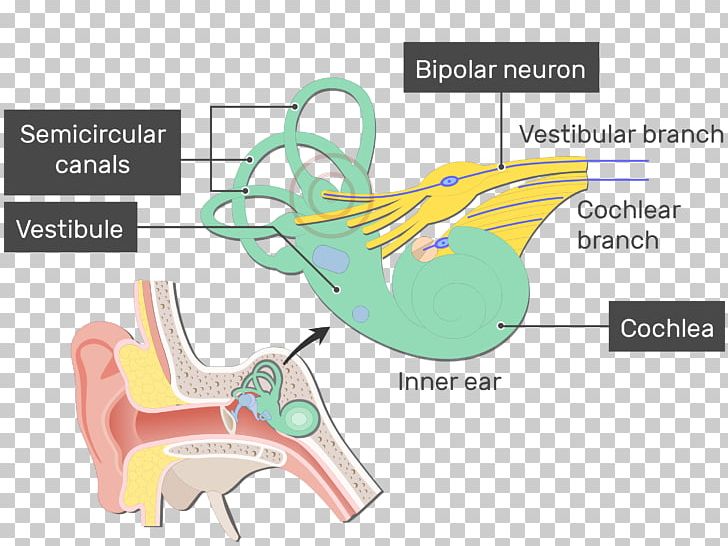
Ear Diagram Vestibule - Human Anatomy
Best chapter 4 adaptive follow-up Flashcards - Quizlet Art-labeling Activity: Neuron structure PICTURE Reading Quiz - Chapter 4 Question 7 Which connective tissue type is found in the walls of large blood vessels and in ligaments supporting transitional epithelia? a) dense irregular connective tissue proper b) elastic connective tissue proper c) adipose connective tissue proper
Post a Comment for "42 art-labeling activity: neuron structure"